

Computational Biology
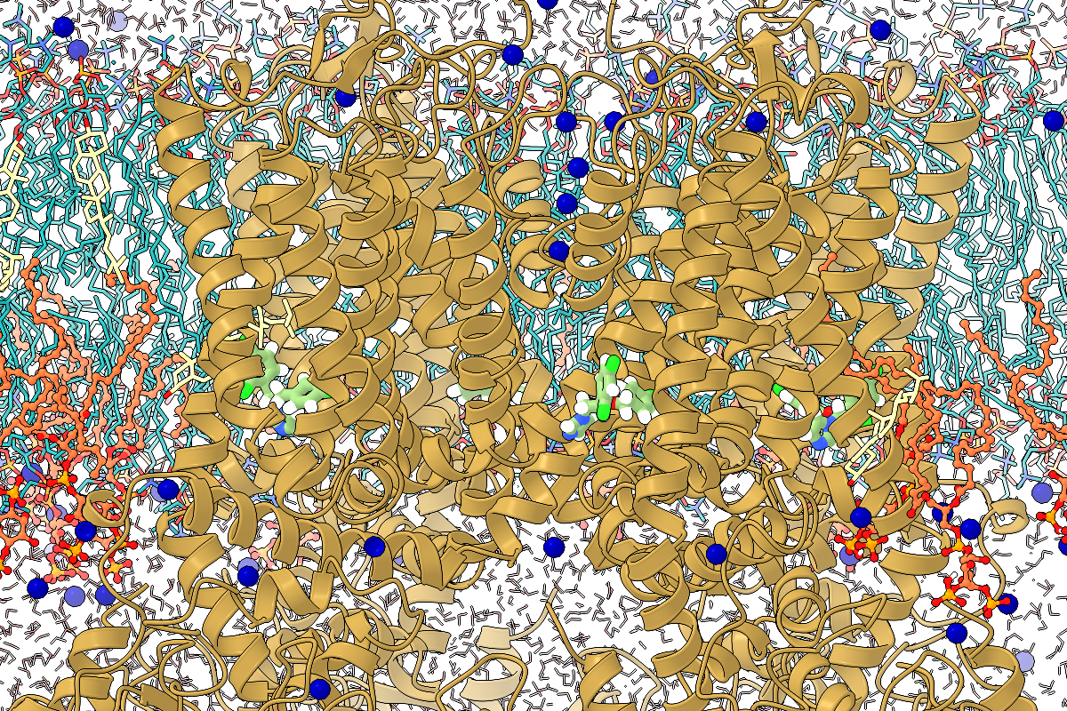
We seek to understand complex biological processes and inform the development of new drugs and proteins based on computer models of biomolecules’ structure and dynamics. Major foci include using simulations to understand proteins’ moving parts and using deep learning to design new proteins and drugs.
Researchers

How are biological structures formed and maintained? How do they manifest biological function? These are central questions in biology that date back thousands of years, to the time of Aristotle. We address the same fundamental questions at a molecular level: how is the diverse and beautiful array of protein and nucleic acid structures formed, and how do they carry out their biological function? Our research addresses these questions using the tools and principles of biophysics, including thermodynamics, statistical mechanics, simulations, and computational geometry.
Computational Biology Computational Biology

The Bowman lab takes a physics-based approach to precision medicine, devising new ways to develop therapeutics and interpret genetic variation by understanding/exploiting protein dynamics. They achieve this using a combination of biophysical experiments, machine learning, physics-based simulations, and the world’s largest distributed computer.
Computational Biology Computational Biology

Our research interests lie at the interface of chemical physics and molecular biology. Our goal is to provide molecular level characterization of complex biomolecular systems and formulate quantitatively accurate microscopic models for predicting the interactions of various therapeutic agents with innate biochemical signaling mechanisms. We employ several computational algorithms ranging from techniques to treat electronic structure, molecular dynamics, Monte Carlo simulations, stochastic kinetic equations, and complex systems analyses in conjunction with the theoretical formalisms of statistical and quantum mechanics, and high performance computing in massively parallel architectures.
Computational Biology Computational Biology

Our goal is to accelerate therapeutic discovery by developing innovative computational approaches that combine physics-based and AI-based methods. We aim to bridge fundamental method development with real-world therapeutic applications, creating more accurate, efficient, and generalizable approaches for drug discovery.
Computational Biology Computational Biology
Related people
Publications
Ward MD, Zimmerman MI, Meller A, Chung M, Swamidass SJ, Bowman GR. Deep learning the structural determinants of protein biochemical properties by comparing structural ensembles with DiffNets. Nat Commun. 2021 May 21;12(1):3023. doi: 10.1038/s41467-021-23246-1. PMID: 34021153; PMCID: PMC8140102.
Abstract
Understanding the structural determinants of a protein's biochemical properties, such as activity and stability, is a major challenge in biology and medicine. Comparing computer simulations of protein variants with different biochemical properties is an increasingly powerful means to drive progress. However, success often hinges on dimensionality reduction algorithms for simplifying the complex ensemble of structures each variant adopts. Unfortunately, common algorithms rely on potentially misleading assumptions about what structural features are important, such as emphasizing larger geometric changes over smaller ones. Here we present DiffNets, self-supervised autoencoders that avoid such assumptions, and automatically identify the relevant features, by requiring that the low-dimensional representations they learn are sufficient to predict the biochemical differences between protein variants. For example, DiffNets automatically identify subtle structural signatures that predict the relative stabilities of β-lactamase variants and duty ratios of myosin isoforms. DiffNets should also be applicable to understanding other perturbations, such as ligand binding.
McShan AC, Flores-Solis D, Sun Y, Garfinkle SE, Toor JS, Young MC, Sgourakis NG. Nat Commun. 2023 Dec 11. doi: 10.1038/s41467-023-43654-9.
Abstract
The conformational landscapes of peptide/human leucocyte antigen (pHLA) protein complexes encompassing tumor neoantigens provide a rationale for target selection towards autologous T cell, vaccine, and antibody-based therapeutic modalities. Here, using complementary biophysical and computational methods, we characterize recurrent RAS55-64 Q61 neoepitopes presented by the common HLA-A*01:01 allotype. We integrate sparse NMR restraints with Rosetta docking to determine the solution structure of NRASQ61K/HLA-A*01:01, which enables modeling of other common RAS55-64 neoepitopes. Hydrogen/deuterium exchange mass spectrometry experiments alongside molecular dynamics simulations reveal differences in solvent accessibility and conformational plasticity across a panel of common Q61 neoepitopes that are relevant for recognition by immunoreceptors. Finally, we predict binding and provide structural models of NRASQ61K antigens spanning the entire HLA allelic landscape, together with in vitro validation for HLA-A*01:191, HLA-B*15:01, and HLA-C*08:02. Our work provides a basis to delineate the solution surface features and immunogenicity of clinically relevant neoepitope/HLA targets for cancer therapy.
Sun Y, Florio TJ, Gupta S, Young MC, Marshall QF, Garfinkle SE, Papadaki GF, Truong HV, Mycek E, Li P, Farrel A, Church NL, Jabar S, Beasley MD, Kiefel BR, Yarmarkovich M, Mallik L, Maris JM, Sgourakis NG. Sci Immunol. 2023 Dec 1. doi: 10.1126/sciimmunol.adj5792.
Abstract
Peptide-centric chimeric antigen receptors (PC-CARs) recognize oncoprotein epitopes displayed by cell-surface human leukocyte antigens (HLAs) and offer a promising strategy for targeted cancer therapy. We have previously developed a PC-CAR targeting a neuroblastoma-associated PHOX2B peptide, leading to robust tumor cell lysis restricted by two common HLA allotypes. Here, we determine the 2.1-angstrom crystal structure of the PC-CAR–PHOX2B–HLA-A*24:02–β2m complex, which reveals the basis for antigen-specific recognition through interactions with CAR complementarity-determining regions (CDRs). This PC-CAR adopts a diagonal docking mode, where interactions with both conserved and polymorphic HLA framework residues permit recognition of multiple HLA allotypes from the A9 serological cross-reactive group, covering a combined global population frequency of up to 46.7%. Biochemical binding assays, molecular dynamics simulations, and structural and functional analyses demonstrate that high-affinity PC-CAR recognition of cross-reactive pHLAs necessitates the presentation of a specific peptide backbone, where subtle structural adaptations of the peptide are critical for high-affinity complex formation, and CAR T cell killing. Our results provide a molecular blueprint for engineering CARs with optimal recognition of tumor-associated antigens in the context of different HLAs, while minimizing cross-reactivity with self-epitopes.
Boby ML, Fearon D, Ferla M, Filep M, Koekemoer L, Robinson MC; COVID Moonshot Consortium‡; Chodera JD, Lee AA, London N, von Delft A, von Delft F, Achdout H, Aimon A, Alonzi DS, Arbon R, Aschenbrenner JC, Balcomb BH, Bar-David E, Barr H, Ben-Shmuel A, Bennett J, Bilenko VA, Borden B, Boulet P, Bowman GR, Brewitz L, Brun J, Bvnbs S, Calmiano M, Carbery A, Carney DW, Cattermole E, Chang E, Chernyshenko E, Clyde A, Coffland JE, Cohen G, Cole JC, Contini A, Cox L, Croll TI, Cvitkovic M, De Jonghe S, Dias A, Donckers K, Dotson DL, Douangamath A, Duberstein S, Dudgeon T, Dunnett LE, Eastman P, Erez N, Eyermann CJ, Fairhead M, Fate G, Fedorov O, Fernandes RS, Ferrins L, Foster R, Foster H, Fraisse L, Gabizon R, García-Sastre A, Gawriljuk VO, Gehrtz P, Gileadi C, Giroud C, Glass WG, Glen RC, Glinert I, Godoy AS, Gorichko M, Gorrie-Stone T, Griffen EJ, Haneef A, Hassell Hart S, Heer J, Henry M, Hill M, Horrell S, Huang QYJ, Huliak VD, Hurley MFD, Israely T, Jajack A, Jansen J, Jnoff E, Jochmans D, John T, Kaminow B, Kang L, Kantsadi AL, Kenny PW, Kiappes JL, Kinakh SO, Kovar B, Krojer T, La VNT, Laghnimi-Hahn S, Lefker BA, Levy H, Lithgo RM, Logvinenko IG, Lukacik P, Macdonald HB, MacLean EM, Makower LL, Malla TR, Marples PG, Matviiuk T, McCorkindale W, McGovern BL, Melamed S, Melnykov KP, Michurin O, Miesen P, Mikolajek H, Milne BF, Minh D, Morris A, Morris GM, Morwitzer MJ, Moustakas D, Mowbray CE, Nakamura AM, Neto JB, Neyts J, Nguyen L, Noske GD, Oleinikovas V, Oliva G, Overheul GJ, Owen CD, Pai R, Pan J, Paran N, Payne AM, Perry B, Pingle M, Pinjari J, Politi B, Powell A, Pšenák V, Pulido I, Puni R, Rangel VL, Reddi RN, Rees P, Reid SP, Reid L, Resnick E, Ripka EG, Robinson RP, Rodriguez-Guerra J, Rosales R, Rufa DA, Saar K, Saikatendu KS, Salah E, Schaller D, Scheen J, Schiffer CA, Schofield CJ, Shafeev M, Shaikh A, Shaqra AM, Shi J, Shurrush K, Singh S, Sittner A, Sjö P, Skyner R, Smalley A, Smeets B, Smilova MD, Solmesky LJ, Spencer J, Strain-Damerell C, Swamy V, Tamir H, Taylor JC, Tennant RE, Thompson W, Thompson A, Tomásio S, Tomlinson CWE, Tsurupa IS, Tumber A, Vakonakis I, van Rij RP, Vangeel L, Varghese FS, Vaschetto M, Vitner EB, Voelz V, Volkamer A, Walsh MA, Ward W, Weatherall C, Weiss S, White KM, Wild CF, Witt KD, Wittmann M, Wright N, Yahalom-Ronen Y, Yilmaz NK, Zaidmann D, Zhang I, Zidane H, Zitzmann N, Zvornicanin SN. Open science discovery of potent noncovalent SARS-CoV-2 main protease inhibitors. Science. 2023 Nov 10;382(6671):eabo7201. doi: 10.1126/science.abo7201. Epub 2023 Nov 10. PMID: 37943932.
Abstract
We report the results of the COVID Moonshot, a fully open-science, crowdsourced, and structure-enabled drug discovery campaign targeting the severe acute respiratory syndrome coronavirus 2 (SARS-CoV-2) main protease. We discovered a noncovalent, nonpeptidic inhibitor scaffold with lead-like properties that is differentiated from current main protease inhibitors. Our approach leveraged crowdsourcing, machine learning, exascale molecular simulations, and high-throughput structural biology and chemistry. We generated a detailed map of the structural plasticity of the SARS-CoV-2 main protease, extensive structure-activity relationships for multiple chemotypes, and a wealth of biochemical activity data. All compound designs (>18,000 designs), crystallographic data (>490 ligand-bound x-ray structures), assay data (>10,000 measurements), and synthesized molecules (>2400 compounds) for this campaign were shared rapidly and openly, creating a rich, open, and intellectual property-free knowledge base for future anticoronavirus drug discovery.
Yarmarkovich M, Marshall QF, Warrington JM, Premaratne R, Farrel A, Groff D, Li W, di Marco M, Runbeck E, Truong H, Toor JS, Tripathi S, Nguyen S, Shen H, Noel T, Church NL, Weiner A, Kendsersky N, Martinez D, Weisberg R, Christie M, Eisenlohr L, Bosse KR, Dimitrov DS, Stevanovic S, Sgourakis NG, Kiefel BR, Maris JM. Nature. 2023 Nov 8. doi: 10.1038/s41586-023-06706-0
Abstract
The majority of oncogenic drivers are intracellular proteins, constraining their immunotherapeutic targeting to mutated peptides (neoantigens) presented by individual human leukocyte antigen (HLA) allotypes1. However, most cancers have a modest mutational burden that is insufficient for generating responses using neoantigen-based therapies2,3. Neuroblastoma is a paediatric cancer that harbours few mutations and is instead driven by epigenetically deregulated transcriptional networks4. Here we show that the neuroblastoma immunopeptidome is enriched with peptides derived from proteins essential for tumorigenesis. We focused on targeting the unmutated peptide QYNPIRTTF discovered on HLA-A*24:02, which is derived from the neuroblastoma-dependency gene and master transcriptional regulator PHOX2B. To target QYNPIRTTF, we developed peptide-centric chimeric antigen receptors (PC-CARs) through a counter panning strategy using predicted potentially cross-reactive peptides. We further proposed that PC-CARs can recognize peptides on additional HLA allotypes when presenting a similar overall molecular surface. Informed by our computational modelling results, we show that PHOX2B PC-CARs also recognize QYNPIRTTF presented by HLA-A*23:01, the most common non-A2 allele in people with African ancestry. Finally, we demonstrate potent and specific killing of neuroblastoma cells expressing these HLAs in vitro and complete tumour regression in mice. These data suggest that PC-CARs have the potential to expand the pool of immunotherapeutic targets to include non-immunogenic intracellular oncoproteins and allow targeting through additional HLA allotypes in a clinical setting.
Gupta S, Nerli S, Kutti Kandy S, Mersky GL, Sgourakis NG. Nat. Commun. 2023 Oct 10. doi: 10.1038/s41467-023-42163-z.
Abstract
The class I proteins of the major histocompatibility complex (MHC-I) display epitopic peptides derived from endogenous proteins on the cell surface for immune surveillance. Accurate modeling of peptides bound to the human MHC, HLA, has been mired by conformational diversity of the central peptide residues, which are critical for recognition by T cell receptors. Here, analysis of X-ray crystal structures within our curated database (HLA3DB) shows that pHLA complexes encompassing multiple HLA allotypes present a discrete set of peptide backbone conformations. Leveraging these backbones, we employ a regression model trained on terms of a physically relevant energy function to develop a comparative modeling approach for nonamer pHLA structures named RepPred. Our method outperforms the top pHLA modeling approach by up to 19% in structural accuracy, and consistently predicts blind targets not included in our training set. Insights from our work may be applied towards predicting antigen immunogenicity, and receptor cross-reactivity.
Ahmed YM, Bowman GR. Phospho-signaling couples polar asymmetry and proteolysis within a membraneless microdomain in C. crescentus. bioRxiv [Preprint]. 2023 Aug 20:2023.08.19.553945. doi: 10.1101/2023.08.19.553945. PMID: 37645878; PMCID: PMC10462113.
Abstract
Asymmetric cell division in bacteria is achieved through cell polarization, where regulatory proteins are directed to specific cell poles. Curiously, both poles contain a membraneless microdomain, established by the polar assembly hub PopZ, through most of the cell cycle, yet many PopZ clients are unipolar and transiently localized. We find that PopZ's interaction with the response regulator CpdR is controlled by phosphorylation, via the histidine kinase CckA. Phosphorylated CpdR does not interact with PopZ and is not localized to cell poles. At poles where CckA acts as a phosphatase, de-phosphorylated CpdR binds directly with PopZ and subsequently recruits ClpX, substrates, and other members of a protease complex to the cell pole. We also find that co-recruitment of protease components and substrates to polar microdomains enhances their coordinated activity. This study connects phosphosignaling with polar assembly and the activity of a protease that triggers cell cycle progression and cell differentiation.
Voelz VA, Pande VS, Bowman GR. Folding@home: Achievements from over 20 years of citizen science herald the exascale era. Biophys J. 2023 Jul 25;122(14):2852-2863. doi: 10.1016/j.bpj.2023.03.028. Epub 2023 Mar 21. PMID: 36945779; PMCID: PMC10398258.
Abstract
Simulations of biomolecules
Simulations of biomolecules have enormous potential to inform our understanding of biology but require extremely demanding calculations. For over 20 years, the Folding@home distributed computing project has pioneered a massively parallel approach to biomolecular simulation, harnessing the resources of citizen scientists across the globe. Here, we summarize the scientific and technical advances this perspective has enabled. As the project's name implies, the early years of Folding@home focused on driving advances in our understanding of protein folding by developing statistical methods for capturing long-timescale processes and facilitating insight into complex dynamical processes. Success laid a foundation for broadening the scope of Folding@home to address other functionally relevant conformational changes, such as receptor signaling, enzyme dynamics, and ligand binding. Continued algorithmic advances, hardware developments such as graphics processing unit (GPU)-based computing, and the growing scale of Folding@home have enabled the project to focus on new areas where massively parallel sampling can be impactful. While previous work sought to expand toward larger proteins with slower conformational changes, new work focuses on large-scale comparative studies of different protein sequences and chemical compounds to better understand biology and inform the development of small-molecule drugs. Progress on these fronts enabled the community to pivot quickly in response to the COVID-19 pandemic, expanding to become the world's first exascale computer and deploying this massive resource to provide insight into the inner workings of the severe acute respiratory syndrome coronavirus 2 (SARS-CoV-2) virus and aid the development of new antivirals. This success provides a glimpse of what is to come as exascale supercomputers come online and as Folding@home continues its work.
Lehman SJ, Meller A, Solieva SO, Lotthammer JM, Greenberg L, Langer SJ, Greenberg MJ, Tardiff JC, Bowman GR, Leinwand L. Divergent Molecular Phenotypes in Point Mutations at the Same Residue in Beta-Myosin Heavy Chain Lead to Distinct Cardiomyopathies. bioRxiv [Preprint]. 2023 Jul 3:2023.07.03.547580. doi: 10.1101/2023.07.03.547580. PMID: 37461648; PMCID: PMC10349964.
Abstract
In genetic cardiomyopathies,
In genetic cardiomyopathies, a frequently described phenomenon is how similar mutations in one protein can lead to discrete clinical phenotypes. One example is illustrated by two mutations in beta myosin heavy chain (β-MHC) that are linked to hypertrophic cardiomyopathy (HCM) (Ile467Val, I467V) and left ventricular non-compaction (LVNC) (Ile467Thr, I467T). To investigate how these missense mutations lead to independent diseases, we studied the molecular effects of each mutation using recombinant human β-MHC Subfragment 1 (S1) in in vitro assays. Both HCM-I467V and LVNC-I467T S1 mutations exhibited similar mechanochemical function, including unchanged ATPase and enhanced actin velocity but had opposing effects on the super-relaxed (SRX) state of myosin. HCM-I467V S1 showed a small reduction in the SRX state, shifting myosin to a more actin-available state that may lead to the "gain-of-function" phenotype commonly described in HCM. In contrast, LVNC-I467T significantly increased the population of myosin in the ultra-slow SRX state. Interestingly, molecular dynamics simulations reveal that I467T allosterically disrupts interactions between ADP and the nucleotide-binding pocket, which may result in an increased ADP release rate. This predicted change in ADP release rate may define the enhanced actin velocity measured in LVNC-I467T, but also describe the uncoupled mechanochemical function for this mutation where the enhanced ADP release rate may be sufficient to offset the increased SRX population of myosin. These contrasting molecular effects may lead to contractile dysregulation that initiates LVNC-associated signaling pathways that progress the phenotype. Together, analysis of these mutations provides evidence that phenotypic complexity originates at the molecular level and is critical to understanding disease progression and developing therapies.
Sun Y, Young MC, Woodward CH, Danon JN, Truong HV, Gupta S, Winters TJ, Font-Burgada J, Burslem GM, Sgourakis NG. Proc. Natl. Acad. Sci. U.S.A. 2023 Jun 20. doi: 10.1073/pnas.2304055120.
Abstract
The polymorphic nature and intrinsic instability of class I major histocompatibility complex (MHC-I) and MHC-like molecules loaded with suboptimal peptides, metabolites, or glycolipids presents a fundamental challenge for identifying disease-relevant antigens and antigen-specific T cell receptors (TCRs), hindering the development of autologous therapeutics. Here, we leverage the positive allosteric coupling between the peptide and light chain (β2 microglobulin, β2m) subunits for binding to the MHC-I heavy chain (HC) through an engineered disulfide bond bridging conserved epitopes across the HC/β2m interface, to generate conformationally stable, peptide-receptive molecules named "open MHC-I." Biophysical characterization shows that open MHC-I molecules are properly folded protein complexes of enhanced thermal stability compared to the wild type when loaded with low- to moderate-affinity peptides. Using solution NMR, we characterize the effects of the disulfide bond on the conformation and dynamics of the MHC-I structure, ranging from local changes in β2m-interacting sites of the peptide-binding groove to long-range effects on the α2-1 helix and α3 domain. The interchain disulfide bond stabilizes MHC-I molecules in an open conformation to promote peptide exchange across multiple human leukocyte antigen (HLA) allotypes, covering representatives from five HLA-A supertypes, six HLA-B supertypes, and oligomorphic HLA-Ib molecules. Our structure-guided design, combined with conditional β-peptide ligands, provides a universal platform to generate ready-to-load MHC-I systems of enhanced stability, enabling a range of approaches to screen antigenic epitope libraries and probe polyclonal TCR repertoires covering highly polymorphic HLA-I allotypes, as well as oligomorphic nonclassical molecules.
Bowman GR, Cox SJ, Dellago C, DuBay KH, Eaves JD, Fletcher DA, Frechette LB, Grünwald M, Klymko K, Ku J, Omar AK, Rabani E, Reichman DR, Rogers JR, Rosnik AM, Rotskoff GM, Schneider AR, Schwierz N, Sivak DA, Vaikuntanathan S, Whitelam S, Widmer-Cooper A. Remembering the Work of Phillip L. Geissler: A Coda to His Scientific Trajectory. Annu Rev Phys Chem. 2023 Apr 24;74:1-27. doi: 10.1146/annurev-physchem-101422-030127. Epub 2023 Jan 31. PMID: 36719975.
Abstract
Phillip L. Geissler made important contributions to the statistical mechanics of biological polymers, heterogeneous materials, and chemical dynamics in aqueous environments. He devised analytical and computational methods that revealed the underlying organization of complex systems at the frontiers of biology, chemistry, and materials science. In this retrospective we celebrate his work at these frontiers.
Meller A, De Oliveira S, Davtyan A, Abramyan T, Bowman GR, van den Bedem H. Discovery of a cryptic pocket in the AI-predicted structure of PPM1D phosphatase explains the binding site and potency of its allosteric inhibitors. Front Mol Biosci. 2023 Apr 18;10:1171143. doi: 10.3389/fmolb.2023.1171143. PMID: 37143823; PMCID: PMC10151774.
Abstract
Virtual screening is a widely used tool for drug discovery, but its predictive power can vary dramatically depending on how much structural data is available. In the best case, crystal structures of a ligand-bound protein can help find more potent ligands. However, virtual screens tend to be less predictive when only ligand-free crystal structures are available, and even less predictive if a homology model or other predicted structure must be used. Here, we explore the possibility that this situation can be improved by better accounting for protein dynamics, as simulations started from a single structure have a reasonable chance of sampling nearby structures that are more compatible with ligand binding. As a specific example, we consider the cancer drug target PPM1D/Wip1 phosphatase, a protein that lacks crystal structures. High-throughput screens have led to the discovery of several allosteric inhibitors of PPM1D, but their binding mode remains unknown. To enable further drug discovery efforts, we assessed the predictive power of an AlphaFold-predicted structure of PPM1D and a Markov state model (MSM) built from molecular dynamics simulations initiated from that structure. Our simulations reveal a cryptic pocket at the interface between two important structural elements, the flap and hinge regions. Using deep learning to predict the pose quality of each docked compound for the active site and cryptic pocket suggests that the inhibitors strongly prefer binding to the cryptic pocket, consistent with their allosteric effect. The predicted affinities for the dynamically uncovered cryptic pocket also recapitulate the relative potencies of the compounds (τb = 0.70) better than the predicted affinities for the static AlphaFold-predicted structure (τb = 0.42). Taken together, these results suggest that targeting the cryptic pocket is a good strategy for drugging PPM1D and, more generally, that conformations selected from simulation can improve virtual screening when limited structural data is available.
Meller A, de Oliveira S, Davtyan A, Abramyan T, Bowman GR, van den Bedem H. Discovery of a cryptic pocket in the AI-predicted structure of PPM1D phosphatase explains the binding site and potency of its allosteric inhibitors. bioRxiv [Preprint]. 2023 Mar 24:2023.03.22.533829. doi: 10.1101/2023.03.22.533829. Update in: Front Mol Biosci. 2023 Apr 18;10:1171143. PMID: 36993233; PMCID: PMC10055338.
Abstract
Virtual screening is a widely used tool for drug discovery, but its predictive power can vary dramatically depending on how much structural data is available. In the best case, crystal structures of a ligand-bound protein can help find more potent ligands. However, virtual screens tend to be less predictive when only ligand-free crystal structures are available, and even less predictive if a homology model or other predicted structure must be used. Here, we explore the possibility that this situation can be improved by better accounting for protein dynamics, as simulations started from a single structure have a reasonable chance of sampling nearby structures that are more compatible with ligand binding. As a specific example, we consider the cancer drug target PPM1D/Wip1 phosphatase, a protein that lacks crystal structures. High-throughput screens have led to the discovery of several allosteric inhibitors of PPM1D, but their binding mode remains unknown. To enable further drug discovery efforts, we assessed the predictive power of an AlphaFold-predicted structure of PPM1D and a Markov state model (MSM) built from molecular dynamics simulations initiated from that structure. Our simulations reveal a cryptic pocket at the interface between two important structural elements, the flap and hinge regions. Using deep learning to predict the pose quality of each docked compound for the active site and cryptic pocket suggests that the inhibitors strongly prefer binding to the cryptic pocket, consistent with their allosteric effect. The predicted affinities for the dynamically uncovered cryptic pocket also recapitulate the relative potencies of the compounds (τ b =0.70) better than the predicted affinities for the static AlphaFold-predicted structure (τ b =0.42). Taken together, these results suggest that targeting the cryptic pocket is a good strategy for drugging PPM1D and, more generally, that conformations selected from simulation can improve virtual screening when limited structural data is available.
Meller A, Bhakat S, Solieva S, Bowman GR. Accelerating Cryptic Pocket Discovery Using AlphaFold. J Chem Theory Comput. 2023 Jul 25;19(14):4355-4363. doi: 10.1021/acs.jctc.2c01189. Epub 2023 Mar 22. PMID: 36948209; PMCID: PMC10373493.
Abstract
Cryptic pockets, or
Cryptic pockets, or pockets absent in ligand-free, experimentally determined structures, hold great potential as drug targets. However, cryptic pocket openings are often beyond the reach of conventional biomolecular simulations because certain cryptic pocket openings involve slow motions. Here, we investigate whether AlphaFold can be used to accelerate cryptic pocket discovery either by generating structures with open pockets directly or generating structures with partially open pockets that can be used as starting points for simulations. We use AlphaFold to generate ensembles for 10 known cryptic pocket examples, including five that were deposited after AlphaFold's training data were extracted from the PDB. We find that in 6 out of 10 cases AlphaFold samples the open state. For plasmepsin II, an aspartic protease from the causative agent of malaria, AlphaFold only captures a partial pocket opening. As a result, we ran simulations from an ensemble of AlphaFold-generated structures and show that this strategy samples cryptic pocket opening, even though an equivalent amount of simulations launched from a ligand-free experimental structure fails to do so. Markov state models (MSMs) constructed from the AlphaFold-seeded simulations quickly yield a free energy landscape of cryptic pocket opening that is in good agreement with the same landscape generated with well-tempered metadynamics. Taken together, our results demonstrate that AlphaFold has a useful role to play in cryptic pocket discovery but that many cryptic pockets may remain difficult to sample using AlphaFold alone.
Voelz VA, Pande VS, Bowman GR. Folding@home: achievements from over twenty years of citizen science herald the exascale era. ArXiv [Preprint]. 2023 Mar 15:arXiv:2303.08993v1. Update in: Biophys J. 2023 Mar 21;: PMID: 36994157; PMCID: PMC10055475.
Abstract
Simulations of biomolecules have enormous potential to inform our understanding of biology but require extremely demanding calculations. For over twenty years, the Folding@home distributed computing project has pioneered a massively parallel approach to biomolecular simulation, harnessing the resources of citizen scientists across the globe. Here, we summarize the scientific and technical advances this perspective has enabled. As the project's name implies, the early years of Folding@home focused on driving advances in our understanding of protein folding by developing statistical methods for capturing long-timescale processes and facilitating insight into complex dynamical processes. Success laid a foundation for broadening the scope of Folding@home to address other functionally relevant conformational changes, such as receptor signaling, enzyme dynamics, and ligand binding. Continued algorithmic advances, hardware developments such as GPU-based computing, and the growing scale of Folding@home have enabled the project to focus on new areas where massively parallel sampling can be impactful. While previous work sought to expand toward larger proteins with slower conformational changes, new work focuses on large-scale comparative studies of different protein sequences and chemical compounds to better understand biology and inform the development of small molecule drugs. Progress on these fronts enabled the community to pivot quickly in response to the COVID-19 pandemic, expanding to become the world's first exascale computer and deploying this massive resource to provide insight into the inner workings of the SARS-CoV-2 virus and aid the development of new antivirals. This success provides a glimpse of what's to come as exascale supercomputers come online, and Folding@home continues its work.
Meller A, Ward M, Borowsky J, Kshirsagar M, Lotthammer JM, Oviedo F, Ferres JL, Bowman GR. Predicting locations of cryptic pockets from single protein structures using the PocketMiner graph neural network. Nat Commun. 2023 Mar 1;14(1):1177. doi: 10.1038/s41467-023-36699-3. PMID: 36859488; PMCID: PMC9977097.
Abstract
Cryptic pockets expand the scope of drug discovery by enabling targeting of proteins currently considered undruggable because they lack pockets in their ground state structures. However, identifying cryptic pockets is labor-intensive and slow. The ability to accurately and rapidly predict if and where cryptic pockets are likely to form from a structure would greatly accelerate the search for druggable pockets. Here, we present PocketMiner, a graph neural network trained to predict where pockets are likely to open in molecular dynamics simulations. Applying PocketMiner to single structures from a newly curated dataset of 39 experimentally confirmed cryptic pockets demonstrates that it accurately identifies cryptic pockets (ROC-AUC: 0.87) >1,000-fold faster than existing methods. We apply PocketMiner across the human proteome and show that predicted pockets open in simulations, suggesting that over half of proteins thought to lack pockets based on available structures likely contain cryptic pockets, vastly expanding the potentially druggable proteome.
Papadaki GF, Ani O, Florio TJ, Young MC, Danon JN, Sun Y, Dersh D, Sgourakis NG. Front. Immunol. 2023 Jan 25. doi: 10.3389/fimmu.2023.1116906.
Major Histocompatibility Complex class I (MHC-I) molecules display self, viral or aberrant epitopic peptides to T cell receptors (TCRs), which employ interactions between complementarity-determining regions with both peptide and MHC-I heavy chain ‘framework’ residues to recognize specific Human Leucocyte Antigens (HLAs). The highly polymorphic nature of the HLA peptide-binding groove suggests a malleability of interactions within a common structural scaffold. Here, using structural data from peptide:MHC-I and pMHC:TCR structures, we first identify residues important for peptide and/or TCR binding. We then outline a fixed-backbone computational design approach for engineering synthetic molecules that combine peptide binding and TCR recognition surfaces from existing HLA allotypes. X-ray crystallography demonstrates that chimeric molecules bridging divergent HLA alleles can bind selected peptide antigens in a specified backbone conformation. Finally, in vitro tetramer staining and biophysical binding experiments using chimeric pMHC-I molecules presenting established antigens further demonstrate the requirement of TCR recognition on interactions with HLA framework residues, as opposed to interactions with peptide-centric Chimeric Antigen Receptors (CARs). Our results underscore a novel, structure-guided platform for developing synthetic HLA molecules with desired properties as screening probes for peptide-centric interactions with TCRs and other therapeutic modalities.
Sun Y, Papadaki GF, Devlin CA, Danon JN, Young MC, Winters TJ, Burslem GM, Procko E, Sgourakis NG, Sci. Adv. 2023 Feb 24. doi: 10.1126/sciadv.ade7151.
Abstract
Immunological chaperones tapasin and TAP binding protein, related (TAPBPR) play key roles in antigenic peptide optimization and quality control of nascent class I major histocompatibility complex (MHC-I) molecules. The polymorphic nature of MHC-I proteins leads to a range of allelic dependencies on chaperones for assembly and cell-surface expression, limiting chaperone-mediated peptide exchange to a restricted set of human leukocyte antigen (HLA) allotypes. Here, we demonstrate and characterize xeno interactions between a chicken TAPBPR ortholog and a complementary repertoire of HLA allotypes, relative to its human counterpart. We find that TAPBPR orthologs recognize empty MHC-I with broader allele specificity and facilitate peptide exchange by maintaining a reservoir of receptive molecules. Deep mutational scanning of human TAPBPR further identifies gain-of-function mutants, resembling the chicken sequence, which can enhance HLA-A*01:01 expression in situ and promote peptide exchange in vitro. These results highlight that polymorphic sites on MHC-I and chaperone surfaces can be engineered to manipulate their interactions, enabling chaperone-mediated peptide exchange on disease-relevant HLA alleles.
Stuchell-Brereton MD, Zimmerman MI, Miller JJ, Mallimadugula UL, Incicco JJ, Roy D, Smith LG, Cubuk J, Baban B, DeKoster GT, Frieden C, Bowman GR, Soranno A. Apolipoprotein E4 has extensive conformational heterogeneity in lipid-free and lipid-bound forms. Proc Natl Acad Sci U S A. 2023 Feb 14;120(7):e2215371120. doi: 10.1073/pnas.2215371120. Epub 2023 Feb 7. PMID: 36749730; PMCID: PMC9963066.
Abstract
The ε4-allele variant of apolipoprotein E (ApoE4) is the strongest genetic risk factor for Alzheimer's disease, although it only differs from its neutral counterpart ApoE3 by a single amino acid substitution. While ApoE4 influences the formation of plaques and neurofibrillary tangles, the structural determinants of pathogenicity remain undetermined due to limited structural information. Previous studies have led to conflicting models of the C-terminal region positioning with respect to the N-terminal domain across isoforms largely because the data are potentially confounded by the presence of heterogeneous oligomers. Here, we apply a combination of single-molecule spectroscopy and molecular dynamics simulations to construct an atomically detailed model of monomeric ApoE4 and probe the effect of lipid association. Importantly, our approach overcomes previous limitations by allowing us to work at picomolar concentrations where only the monomer is present. Our data reveal that ApoE4 is far more disordered and extended than previously thought and retains significant conformational heterogeneity after binding lipids. Comparing the proximity of the N- and C-terminal domains across the three major isoforms (ApoE4, ApoE3, and ApoE2) suggests that all maintain heterogeneous conformations in their monomeric form, with ApoE2 adopting a slightly more compact ensemble. Overall, these data provide a foundation for understanding how ApoE4 differs from nonpathogenic and protective variants of the protein.
Meller A, Lotthammer JM, Smith LG, Novak B, Lee LA, Kuhn CC, Greenberg L, Leinwand LA, Greenberg MJ, Bowman GR. Drug specificity and affinity are encoded in the probability of cryptic pocket opening in myosin motor domains. Elife. 2023 Jan 27;12:e83602. doi: 10.7554/eLife.83602. PMID: 36705568; PMCID: PMC9995120.
Abstract
The design of compounds that can discriminate between closely related target proteins remains a central challenge in drug discovery. Specific therapeutics targeting the highly conserved myosin motor family are urgently needed as mutations in at least six of its members cause numerous diseases. Allosteric modulators, like the myosin-II inhibitor blebbistatin, are a promising means to achieve specificity. However, it remains unclear why blebbistatin inhibits myosin-II motors with different potencies given that it binds at a highly conserved pocket that is always closed in blebbistatin-free experimental structures. We hypothesized that the probability of pocket opening is an important determinant of the potency of compounds like blebbistatin. To test this hypothesis, we used Markov state models (MSMs) built from over 2 ms of aggregate molecular dynamics simulations with explicit solvent. We find that blebbistatin's binding pocket readily opens in simulations of blebbistatin-sensitive myosin isoforms. Comparing these conformational ensembles reveals that the probability of pocket opening correctly identifies which isoforms are most sensitive to blebbistatin inhibition and that docking against MSMs quantitatively predicts blebbistatin binding affinities (R2=0.82). In a blind prediction for an isoform (Myh7b) whose blebbistatin sensitivity was unknown, we find good agreement between predicted and measured IC50s (0.67 μM vs. 0.36 μM). Therefore, we expect this framework to be useful for the development of novel specific drugs across numerous protein targets.
Tamadonfar KO, Di Venanzio G, Pinkner JS, Dodson KW, Kalas V, Zimmerman MI, Bazan Villicana J, Bowman GR, Feldman MF, Hultgren SJ. Structure-function correlates of fibrinogen binding by Acinetobacter adhesins critical in catheter-associated urinary tract infections. Proc Natl Acad Sci U S A. 2023 Jan 24;120(4):e2212694120. doi: 10.1073/pnas.2212694120. Epub 2023 Jan 18. PMID: 36652481; PMCID: PMC9942807.
Abstract
Multidrug-resistant
Multidrug-resistant Acinetobacter baumannii infections are an urgent clinical problem and can cause difficult-to-treat nosocomial infections. During such infections, like catheter-associated urinary tract infections (CAUTI), A. baumannii rely on adhesive, extracellular fibers, called chaperone-usher pathway (CUP) pili for critical binding interactions. The A. baumannii uropathogenic strain, UPAB1, and the pan-European subclone II isolate, ACICU, use the CUP pili Abp1 and Abp2 (previously termed Cup and Prp, respectively) in tandem to establish CAUTIs, specifically to facilitate bacterial adherence and biofilm formation on the implanted catheter. Abp1 and Abp2 pili are tipped with two domain tip adhesins, Abp1D and Abp2D, respectively. We discovered that both adhesins bind fibrinogen, a critical host wound response protein that is released into the bladder upon catheterization and is subsequently deposited on the catheter. The crystal structures of the Abp1D and Abp2D receptor-binding domains were determined and revealed that they both contain a large, distally oriented pocket, which mediates binding to fibrinogen and other glycoproteins. Genetic, biochemical, and biophysical studies revealed that interactions with host proteins are governed by several critical residues in and along the edge of the binding pocket, one of which regulates the structural stability of an anterior loop motif. K34, located outside of the pocket but interacting with the anterior loop, also regulates the binding affinity of the protein. This study illuminates the mechanistic basis of the critical fibrinogen-coated catheter colonization step in A. baumannii CAUTI pathogenesis.
Agard DA, Bowman GR, DeGrado W, Dokholyan NV, Zhou HX. Solution of the protein structure prediction problem at last: crucial innovations and next frontiers. Fac Rev. 2022 Dec 14;11:38. doi: 10.12703/r-01-0000020. PMID: 36644294; PMCID: PMC9815721.
Abstract
The protein structure
The protein structure prediction problem is solved, at last, thanks in large part to the use of artificial intelligence. The structures predicted by AlphaFold and RoseTTAFold are becoming the requisite starting point for many protein scientists. New frontiers, such as the conformational sampling of intrinsically disordered proteins, are emerging.
Knapp BD, Ward MD, Bowman GR, Shi H, Huang KC. Multiple conserved states characterize the twist landscape of the bacterial actin homolog MreB. Comput Struct Biotechnol J. 2022 Oct 7;20:5838-5846. doi: 10.1016/j.csbj.2022.10.008. PMID: 36382191; PMCID: PMC9627593.
Abstract
Filament formation by cytoskeletal proteins is critical to their involvement in myriad cellular processes. The bacterial actin homolog MreB, which is essential for cell-shape determination in many rod-shaped bacteria, has served as a model system for studying the mechanics of cytoskeletal filaments. Previous molecular dynamics (MD) simulations revealed that the twist of MreB double protofilaments is dependent on the bound nucleotide, as well as binding to the membrane or the accessory protein RodZ, and MreB mutations that modulate twist also affect MreB spatial organization and cell shape. Here, we show that MreB double protofilaments can adopt multiple twist states during microsecond-scale MD simulations. A deep learning algorithm trained only on high- and low-twist states robustly identified all twist conformations across most perturbations of ATP-bound MreB, suggesting the existence of a conserved set of states whose occupancy is affected by each perturbation to MreB. Simulations replacing ATP with ADP indicated that twist states were generally stable after hydrolysis. These findings suggest a rich twist landscape that could provide the capacity to tune MreB activity and therefore its effects on cell shape.
Polino AJ, Miller JJ, Bhakat S, Mukherjee S, Bobba S, Bowman GR, Goldberg DE. The nepenthesin insert in the Plasmodium falciparum aspartic protease plasmepsin V is necessary for enzyme function. J Biol Chem. 2022 Sep;298(9):102355. doi: 10.1016/j.jbc.2022.102355. Epub 2022 Aug 9. PMID: 35952758; PMCID: PMC9478907.
Abstract
Plasmepsin V (PM
Plasmepsin V (PM V) is a pepsin-like aspartic protease essential for growth of the malarial parasite Plasmodium falciparum. Previous work has shown PM V to be an endoplasmic reticulum-resident protease that processes parasite proteins destined for export into the host cell. Depletion or inhibition of the enzyme is lethal during asexual replication within red blood cells as well as during the formation of sexual stage gametocytes. The structure of the Plasmodium vivax PM V has been characterized by X-ray crystallography, revealing a canonical pepsin fold punctuated by structural features uncommon to secretory aspartic proteases; however, the function of this unique structure is unclear. Here, we used parasite genetics to probe these structural features by attempting to rescue lethal PM V depletion with various mutant enzymes. We found an unusual nepenthesin 1-type insert in the PM V gene to be essential for parasite growth and PM V activity. Mutagenesis of the nepenthesin insert suggests that both its amino acid sequence and one of the two disulfide bonds that undergird its structure are required for the insert's role in PM V function. Furthermore, molecular dynamics simulations paired with Markov state modeling suggest that mutations to the nepenthesin insert may allosterically affect PM V catalysis through multiple mechanisms. Taken together, these data provide further insights into the structure of the P. falciparum PM V protease.
Huang Z, Liu H, Nix J, Xu R, Knoverek CR, Bowman GR, Amarasinghe GK, Sibley LD. The intrinsically disordered protein TgIST from Toxoplasma gondii inhibits STAT1 signaling by blocking cofactor recruitment. Nat Commun. 2022 Jul 13;13(1):4047. doi: 10.1038/s41467-022-31720-7. PMID: 35831295; PMCID: PMC9279507.
Abstract
Signal transducer and activator of transcription (STAT) proteins communicate from cell-surface receptors to drive transcription of immune response genes. The parasite Toxoplasma gondii blocks STAT1-mediated gene expression by secreting the intrinsically disordered protein TgIST that traffics to the host nucleus, binds phosphorylated STAT1 dimers, and occupies nascent transcription sites that unexpectedly remain silenced. Here we define a core region within internal repeats of TgIST that is necessary and sufficient to block STAT1-mediated gene expression. Cellular, biochemical, mutational, and structural data demonstrate that the repeat region of TgIST adopts a helical conformation upon binding to STAT1 dimers. The binding interface is defined by a groove formed from two loops in the STAT1 SH2 domains that reorient during dimerization. TgIST binding to this newly exposed site at the STAT1 dimer interface alters its conformation and prevents the recruitment of co-transcriptional activators, thus defining the mechanism of blocked transcription.
Liu C, Karabina A, Meller A, Bhattacharjee A, Agostino CJ, Bowman GR, Ruppel KM, Spudich JA, Leinwand LA. Homologous mutations in β, embryonic, and perinatal muscle myosins have divergent effects on molecular power generation. bioRxiv [Preprint]. 2023 Jul 2:2023.07.02.547385. doi: 10.1101/2023.07.02.547385. PMID: 37425764; PMCID: PMC10327197.
Abstract
Mutations at a highly conserved homologous residue in three closely related muscle myosins cause three distinct diseases involving muscle defects: R671C in β-cardiac myosin causes hypertrophic cardiomyopathy, R672C and R672H in embryonic skeletal myosin cause Freeman Sheldon syndrome, and R674Q in perinatal skeletal myosin causes trismus-pseudocamptodactyly syndrome. It is not known if their effects at the molecular level are similar to one another or correlate with disease phenotype and severity. To this end, we investigated the effects of the homologous mutations on key factors of molecular power production using recombinantly expressed human β, embryonic, and perinatal myosin subfragment-1. We found large effects in the developmental myosins, with the most dramatic in perinatal, but minimal effects in β myosin, and magnitude of changes correlated partially with clinical severity. The mutations in the developmental myosins dramatically decreased the step size and load-sensitive actin-detachment rate of single molecules measured by optical tweezers, in addition to decreasing ATPase cycle rate. In contrast, the only measured effect of R671C in β myosin was a larger step size. Our measurements of step size and bound times predicted velocities consistent with those measured in an in vitro motility assay. Finally, molecular dynamics simulations predicted that the arginine to cysteine mutation in embryonic, but not β, myosin may reduce pre-powerstroke lever arm priming and ADP pocket opening, providing a possible structural mechanism consistent with the experimental observations. This paper presents the first direct comparisons of homologous mutations in several different myosin isoforms, whose divergent functional effects are yet another testament to myosin's highly allosteric nature.
Cruz MA, Frederick TE, Mallimadugula UL, Singh S, Vithani N, Zimmerman MI, Porter JR, Moeder KE, Amarasinghe GK, Bowman GR. A cryptic pocket in Ebola VP35 allosterically controls RNA binding. Nat Commun. 2022 Apr 27;13(1):2269. doi: 10.1038/s41467-022-29927-9. PMID: 35477718; PMCID: PMC9046395.
Abstract
Protein-protein and protein-nucleic
Protein-protein and protein-nucleic acid interactions are often considered difficult drug targets because the surfaces involved lack obvious druggable pockets. Cryptic pockets could present opportunities for targeting these interactions, but identifying and exploiting these pockets remains challenging. Here, we apply a general pipeline for identifying cryptic pockets to the interferon inhibitory domain (IID) of Ebola virus viral protein 35 (VP35). VP35 plays multiple essential roles in Ebola's replication cycle but lacks pockets that present obvious utility for drug design. Using adaptive sampling simulations and machine learning algorithms, we predict VP35 harbors a cryptic pocket that is allosterically coupled to a key dsRNA-binding interface. Thiol labeling experiments corroborate the predicted pocket and mutating the predicted allosteric network supports our model of allostery. Finally, covalent modifications that mimic drug binding allosterically disrupt dsRNA binding that is essential for immune evasion. Based on these results, we expect this pipeline will be applicable to other proteins.
Jansen J, Reimer KC, Nagai JS, Varghese FS, Overheul GJ, de Beer M, Roverts R, Daviran D, Fermin LAS, Willemsen B, Beukenboom M, Djudjaj S, von Stillfried S, van Eijk LE, Mastik M, Bulthuis M, Dunnen WD, van Goor H, Hillebrands JL, Triana SH, Alexandrov T, Timm MC, van den Berge BT, van den Broek M, Nlandu Q, Heijnert J, Bindels EMJ, Hoogenboezem RM, Mooren F, Kuppe C, Miesen P, Grünberg K, Ijzermans T, Steenbergen EJ, Czogalla J, Schreuder MF, Sommerdijk N, Akiva A, Boor P, Puelles VG, Floege J, Huber TB; COVID Moonshot consortium; van Rij RP, Costa IG, Schneider RK, Smeets B, Kramann R. SARS-CoV-2 infects the human kidney and drives fibrosis in kidney organoids. Cell Stem Cell. 2022 Feb 3;29(2):217-231.e8. doi: 10.1016/j.stem.2021.12.010. Epub 2021 Dec 25. PMID: 35032430; PMCID: PMC8709832.
Abstract
Kidney failure is
Kidney failure is frequently observed during and after COVID-19, but it remains elusive whether this is a direct effect of the virus. Here, we report that SARS-CoV-2 directly infects kidney cells and is associated with increased tubule-interstitial kidney fibrosis in patient autopsy samples. To study direct effects of the virus on the kidney independent of systemic effects of COVID-19, we infected human-induced pluripotent stem-cell-derived kidney organoids with SARS-CoV-2. Single-cell RNA sequencing indicated injury and dedifferentiation of infected cells with activation of profibrotic signaling pathways. Importantly, SARS-CoV-2 infection also led to increased collagen 1 protein expression in organoids. A SARS-CoV-2 protease inhibitor was able to ameliorate the infection of kidney cells by SARS-CoV-2. Our results suggest that SARS-CoV-2 can directly infect kidney cells and induce cell injury with subsequent fibrosis. These data could explain both acute kidney injury in COVID-19 patients and the development of chronic kidney disease in long COVID.
Topf M, Rosta E, Bowman GR, Bonomi M. Editorial: Experiments and Simulations: A Pas de Deux to Unravel Biological Function. Front Mol Biosci. 2021 Nov 29;8:799406. doi: 10.3389/fmolb.2021.799406. PMID: 34912853; PMCID: PMC8667856.
Knoverek CR, Mallimadugula UL, Singh S, Rennella E, Frederick TE, Yuwen T, Raavicharla S, Kay LE, Bowman GR. Opening of a cryptic pocket in β-lactamase increases penicillinase activity. Proc Natl Acad Sci U S A. 2021 Nov 23;118(47):e2106473118. doi: 10.1073/pnas.2106473118. PMID: 34799442; PMCID: PMC8617505.
Abstract
Understanding the functional
Understanding the functional role of protein-excited states has important implications in protein design and drug discovery. However, because these states are difficult to find and study, it is still unclear if excited states simply result from thermal fluctuations and generally detract from function or if these states can actually enhance protein function. To investigate this question, we consider excited states in β-lactamases and particularly a subset of states containing a cryptic pocket which forms under the Ω-loop. Given the known importance of the Ω-loop and the presence of this pocket in at least two homologs, we hypothesized that these excited states enhance enzyme activity. Using thiol-labeling assays to probe Ω-loop pocket dynamics and kinetic assays to probe activity, we find that while this pocket is not completely conserved across β-lactamase homologs, those with the Ω-loop pocket have a higher activity against the substrate benzylpenicillin. We also find that this is true for TEM β-lactamase variants with greater open Ω-loop pocket populations. We further investigate the open population using a combination of NMR chemical exchange saturation transfer experiments and molecular dynamics simulations. To test our understanding of the Ω-loop pocket's functional role, we designed mutations to enhance/suppress pocket opening and observed that benzylpenicillin activity is proportional to the probability of pocket opening in our designed variants. The work described here suggests that excited states containing cryptic pockets can be advantageous for function and may be favored by natural selection, increasing the potential utility of such cryptic pockets as drug targets.
Malik M, Ward MD, Fang Y, Porter JR, Zimmerman MI, Koelblen T, Roh M, Frolova AI, Burris TP, Bowman GR, Imoukhuede PI, England SK. Naturally Occurring Genetic Variants in the Oxytocin Receptor Alter Receptor Signaling Profiles. ACS Pharmacol Transl Sci. 2021 Sep 8;4(5):1543-1555. doi: 10.1021/acsptsci.1c00095. PMID: 34661073; PMCID: PMC8506602.
Abstract
The hormone oxytocin
The hormone oxytocin is commonly administered during childbirth to initiate and strengthen uterine contractions and prevent postpartum hemorrhage. However, patients have wide variation in the oxytocin dose required for a clinical response. To begin to uncover the mechanisms underlying this variability, we screened the 11 most prevalent missense genetic variants in the oxytocin receptor (OXTR) gene. We found that five variants, V45L, P108A, L206V, V281M, and E339K, significantly altered oxytocin-induced Ca2+ signaling or β-arrestin recruitment and proceeded to assess the effects of these variants on OXTR trafficking to the cell membrane, desensitization, and internalization. The variants P108A and L206V increased OXTR localization to the cell membrane, whereas V281M and E339K caused OXTR to be retained inside the cell. We examined how the variants altered the balance between OXTR activation and desensitization, which is critical for appropriate oxytocin dosing. The E339K variant impaired OXTR activation, internalization, and desensitization to roughly equal extents. In contrast, V281M decreased OXTR activation but had no effect on internalization and desensitization. V45L and P108A did not alter OXTR activation but did impair β-arrestin recruitment, internalization, and desensitization. Molecular dynamics simulations predicted that V45L and P108A prevent extension of the first intracellular loop of OXTR, thus inhibiting β-arrestin binding. Overall, our data suggest mechanisms by which OXTR genetic variants could alter clinical response to oxytocin.
Vithani N, Ward MD, Zimmerman MI, Novak B, Borowsky JH, Singh S, Bowman GR. SARS-CoV-2 Nsp16 activation mechanism and a cryptic pocket with pan-coronavirus antiviral potential. Biophys J. 2021 Jul 20;120(14):2880-2889. doi: 10.1016/j.bpj.2021.03.024. Epub 2021 Mar 29. PMID: 33794150; PMCID: PMC8007187.
Abstract
Coronaviruses have caused
Coronaviruses have caused multiple epidemics in the past two decades, in addition to the current COVID-19 pandemic that is severely damaging global health and the economy. Coronaviruses employ between 20 and 30 proteins to carry out their viral replication cycle, including infection, immune evasion, and replication. Among these, nonstructural protein 16 (Nsp16), a 2'-O-methyltransferase, plays an essential role in immune evasion. Nsp16 achieves this by mimicking its human homolog, CMTr1, which methylates mRNA to enhance translation efficiency and distinguish self from other. Unlike human CMTr1, Nsp16 requires a binding partner, Nsp10, to activate its enzymatic activity. The requirement of this binding partner presents two questions that we investigate in this manuscript. First, how does Nsp10 activate Nsp16? Although experimentally derived structures of the active Nsp16/Nsp10 complex exist, structures of inactive, monomeric Nsp16 have yet to be solved. Therefore, it is unclear how Nsp10 activates Nsp16. Using over 1 ms of molecular dynamics simulations of both Nsp16 and its complex with Nsp10, we investigate how the presence of Nsp10 shifts Nsp16's conformational ensemble to activate it. Second, guided by this activation mechanism and Markov state models, we investigate whether Nsp16 adopts inactive structures with cryptic pockets that, if targeted with a small molecule, could inhibit Nsp16 by stabilizing its inactive state. After identifying such a pocket in SARS-CoV2 Nsp16, we show that this cryptic pocket also opens in SARS-CoV1 and MERS but not in human CMTr1. Therefore, it may be possible to develop pan-coronavirus antivirals that target this cryptic pocket.
Moreno-Gómez M, Bueno-Marí R, Carr BT, Bowman GR, Faherty GW, Gobbi C, Palm JM, Van Sloun P, Miranda MÁ. Two New Alternatives to the Conventional Arm-in-Cage Test for Assessing Topical Repellents. J Med Entomol. 2021 Jul 16;58(4):1826-1838. doi: 10.1093/jme/tjab050. PMID: 33769523; PMCID: PMC8285018.
Abstract
European guidelines for
European guidelines for testing attractant and repellent efficacy (i.e., Product type 19 [PT19]) have been in revision since 2017. A key topic of discussion is the current approach to evaluating topical repellents. The European Chemical Agency has stated field testing should be avoided because of mosquito-borne disease risks. However, the most common laboratory method, the arm-in-cage (AIC) test, may limit the reliable extrapolation of lab results to field conditions. This study's main goal was to assess alternative laboratory methods for evaluating topical mosquito repellents that use mosquito landing rates more representative of those in the field. The study took place at three European testing labs using 30 study participants per test and the mosquito, Aedes albopictus (Skuse, 1894, Diptera: Culicidae). In phase 1, a conventional AIC test and a sleeved AIC test were performed. Respectively, the arm area exposed was 600 and 100 cm2, and cage volume was 0.040 and 0.064 m3. Mosquito density was the same for both: 1 female/840 cm3. In phase 2, room-based testing (40 ± 5 mosquitoes in 25-30 m3) was used as a proxy for field testing. The mosquito repellent employed was 15% N,N-diethyl-m-toluamide in ethanol at two doses: 1 and 0.5 g/600 cm2. The protection times measured at each laboratory were analyzed both separately and together using nonparametric (Kruskal-Wallis) test. The two alternatives methods showed to be potential alternatives to the current AIC method recreated field mosquito landing rates and achieved reproducible protection times across laboratories.
Zimmerman MI, Porter JR, Ward MD, Singh S, Vithani N, Meller A, Mallimadugula UL, Kuhn CE, Borowsky JH, Wiewiora RP, Hurley MFD, Harbison AM, Fogarty CA, Coffland JE, Fadda E, Voelz VA, Chodera JD, Bowman GR. SARS-CoV-2 simulations go exascale to predict dramatic spike opening and cryptic pockets across the proteome. Nat Chem. 2021 Jul;13(7):651-659. doi: 10.1038/s41557-021-00707-0. Epub 2021 May 24. PMID: 34031561; PMCID: PMC8249329.
Abstract
SARS-CoV-2 has intricate mechanisms for initiating infection, immune evasion/suppression and replication that depend on the structure and dynamics of its constituent proteins. Many protein structures have been solved, but far less is known about their relevant conformational changes. To address this challenge, over a million citizen scientists banded together through the Folding@home distributed computing project to create the first exascale computer and simulate 0.1 seconds of the viral proteome. Our adaptive sampling simulations predict dramatic opening of the apo spike complex, far beyond that seen experimentally, explaining and predicting the existence of 'cryptic' epitopes. Different spike variants modulate the probabilities of open versus closed structures, balancing receptor binding and immune evasion. We also discover dramatic conformational changes across the proteome, which reveal over 50 'cryptic' pockets that expand targeting options for the design of antivirals. All data and models are freely available online, providing a quantitative structural atlas.
Cubuk J, Alston JJ, Incicco JJ, Singh S, Stuchell-Brereton MD, Ward MD, Zimmerman MI, Vithani N, Griffith D, Wagoner JA, Bowman GR, Hall KB, Soranno A, Holehouse AS. The SARS-CoV-2 nucleocapsid protein is dynamic, disordered, and phase separates with RNA. Nat Commun. 2021 Mar 29;12(1):1936. doi: 10.1038/s41467-021-21953-3. PMID: 33782395; PMCID: PMC8007728.
Abstract
The SARS-CoV-2 nucleocapsid
The SARS-CoV-2 nucleocapsid (N) protein is an abundant RNA-binding protein critical for viral genome packaging, yet the molecular details that underlie this process are poorly understood. Here we combine single-molecule spectroscopy with all-atom simulations to uncover the molecular details that contribute to N protein function. N protein contains three dynamic disordered regions that house putative transiently-helical binding motifs. The two folded domains interact minimally such that full-length N protein is a flexible and multivalent RNA-binding protein. N protein also undergoes liquid-liquid phase separation when mixed with RNA, and polymer theory predicts that the same multivalent interactions that drive phase separation also engender RNA compaction. We offer a simple symmetry-breaking model that provides a plausible route through which single-genome condensation preferentially occurs over phase separation, suggesting that phase separation offers a convenient macroscopic readout of a key nanoscopic interaction.