

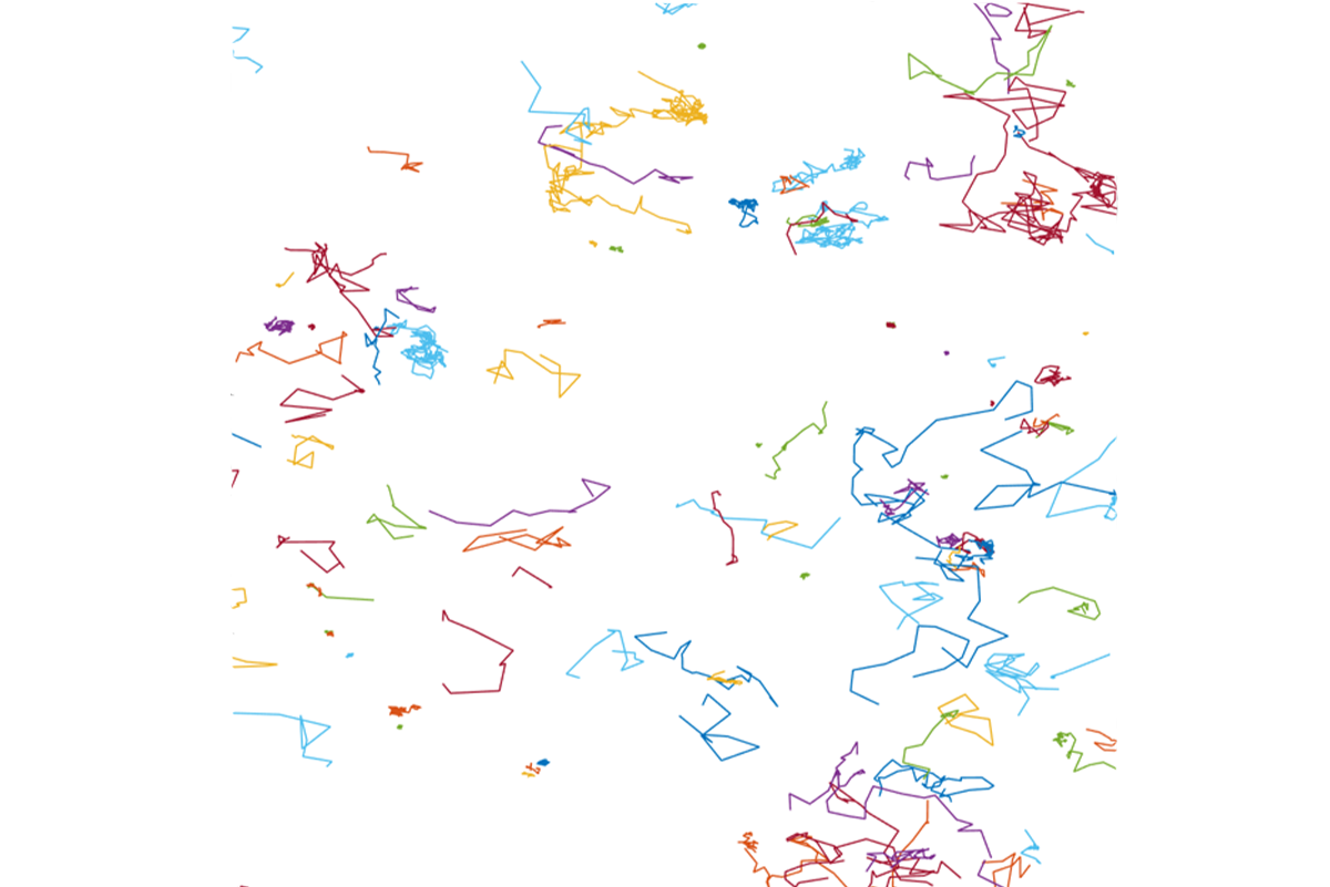
Single Molecule Imaging
Single molecule techniques investigate the complex behaviors of biomolecules in vitro and in cellular environments, revealing their molecular structure, dynamics, interactions, and functions. By avoiding the need for averaging that is inherent in bulk measurements, these powerful methods provide nuanced insights into the diverse nature of molecular activities, enhancing our understanding of molecular heterogeneity.
Researchers





Publications
Abstract
Abstract
Abstract
Proteins can organize into dynamic, functionally important assemblies on fluid membrane surfaces. Phase separation has emerged as an important mechanism for forming such protein assemblies on the membrane during cell signaling, endocytosis, and cytoskeleton regulation. Protein-protein phase separation thus adds novel fluid mosaics to the classical Singer and Nicolson model. Protein condensates formed in this process can modulate membrane morphologies. This is evident from recent reports of protein condensate-driven membrane reshaping in processes such as endocytosis, autophagosome formation, and protein storage vacuole morphogenesis in plants. Lateral phase separation (on the membrane surface) of peripheral curvature coupling proteins can modulate such membrane morphological transitions. Additionally, three-dimensional protein phase separation can result in droplets that through adhesion can affect membrane shape changes. How do these condensate-driven curvature generation mechanisms contrast with the classically recognized scaffolding and amphipathic helix insertion activities of specific membrane remodeling proteins? A salient feature of these condensate-driven membrane activities is that they depend upon both macroscopic features (such as interfacial energies of the condensate, membrane, and cytosol) as well as microscopic, molecular-level interactions (such as protein-lipid binding). This review highlights the current understanding of the mechanisms underlying curvature generation by protein condensates in various biological pathways.
Abstract
Abstract
BAR (Bin/Amphiphysin/Rvs) domain containing proteins function as lipid bilayer benders and curvature sensors, and they contribute to membrane shaping involved in cell signaling and metabolism. The mechanism for their membrane shape sensing has been investigated by both equilibrium binding and kinetic studies. In prior research, stopped-flow spectroscopy has been used to deduce a positive dependence on membrane curvature for the binding rate constant, kon, of a BAR protein called endophilin. However, the impact of bulk diffusion of endophilin, on the kinetic binding parameters has not been thoroughly considered. Employing similar methods, and using lipid vesicles of multiple sizes, we obtained a linear dependence of kon on vesicle curvature. However, we found that the observed relation can be explained without considering the local curvature sensing ability of endophilin in the membrane association process. In contrast, the diffusion-independent unbinding rate constant (koff) obtained from stopped-flow measurements shows a negative dependence on membrane curvature, which is controlled/mediated by endophilin-membrane interactions. This latter dependency, in addition to protein–protein interactions on the membrane, explains the selective binding of BAR proteins to highly curved membranes in equilibrium binding experiments.
Abstract
Lipid bilayer membranes undergo rapid bending undulations with wavelengths from tens of nanometers to tens of microns due to thermal fluctuations. Here, we probe such undulations and the membranes’ mechanics by measuring the time-varying orientation of single gold nanorods (GNRs) adhered to the membrane, using high-speed dark field microscopy. In a lipid vesicle, such measurements allow the determination of the membrane’s viscosity, bending rigidity, and tension as well as the friction coefficient for sliding of the monolayers over one another. The in-plane rotation of the GNR is hindered by undulations in a tension dependent manner, consistent with simulations. The motion of single GNRs adhered to the plasma membrane of living cultured cells similarly reveals the membrane’s complex physics and coupling to the cell’s actomyosin cortex.
Abstract
Abstract
Abstract
The eukaryotic plasma membrane's lipid composition is found to be ubiquitously asymmetric comparing inner and outer leaflets. This membrane lipid asymmetry plays a crucial role in diverse cellular processes critical for cell survival. A specialized set of transmembrane proteins called translocases, or flippases, have evolved to maintain this membrane lipid asymmetry in an energy-dependent manner. One potential consequence of local variations in membrane lipid asymmetry is membrane remodeling, which is essential for cellular processes such as intracellular trafficking. Recently, there has been a surge in the identification and characterization of flippases, which has significantly advanced the understanding of their functional mechanisms. Furthermore, there are intriguing possibilities for a coupling between membrane curvature and flippase activity. In this review we highlight studies that link membrane shape and remodeling to differential stresses generated by the activity of lipid flippases with an emphasis on data obtained through model membrane systems. We review the common mechanistic models of flippase-mediated lipid flipping and discuss common techniques used to test lipid flippase activity. We then compare the existing data on lipid translocation rates by flippases and conclude with potential future directions for this field.
Abstract
Liquid-liquid phase separation has recently emerged as an important fundamental organizational phenomenon in biological settings. Most studies of biological phase separation have focused on droplets that “condense” from solution above a critical concentration, forming so-called “membraneless organelles” suspended in solution. However, membranes are ubiquitous throughout cells, and many biomolecular condensates interact with membrane surfaces. Such membrane-associated phase-separated systems range from clusters of integral or peripheral membrane proteins in the plane of the membrane to free, spherical droplets wetting membrane surfaces to droplets containing small lipid vesicles. In this review, we consider phase-separated liquids that interact with membrane surfaces and we discuss the consequences of those interactions. The physical properties of distinct liquid phases in contact with bilayers can reshape the membrane, and liquid-liquid phase separation can construct membrane-associated protein structures, modulate their function, and organize collections of lipid vesicles dynamically. We summarize the common phenomena that arise in these systems of liquid phases and membranes.
Abstract
Abstract
Phosphatidylinositol-4,5-bisphosphate (PIP2) is an important signaling lipid in eukaryotic cell plasma membranes, playing an essential role in diverse cellular processes. The headgroup of PIP2 is highly negatively charged, and this lipid displays a high critical micellar concentration compared to housekeeping phospholipid analogs. Given the crucial role of PIP2, it is imperative to study its localization, interaction with proteins, and membrane-shaping properties. Biomimetic membranes have served extensively to elucidate structural and functional aspects of cell membranes including protein-lipid and lipid-lipid interactions, as well as membrane mechanics. Incorporation of PIP2 into biomimetic membranes, however, has at times resulted in discrepant findings described in the literature. With the goal to elucidate the mechanical consequences of PIP2 incorporation, we studied the desorption of PIP2 from biomimetic giant unilamellar vesicles by means of a fluorescent marker. A decrease in fluorescence intensity with the age of the vesicles suggested that PIP2 lipids were being desorbed from the outer leaflet of the membrane. To evaluate whether this desorption was asymmetric, the vesicles were systematically diluted. This resulted in an increase in the number of internally tubulated vesicles within minutes after dilution, suggesting that the desorption was asymmetric and also generated membrane curvature. By means of a saturated chain homolog of PIP2, we showed that the fast desorption of PIP2 is facilitated by presence of an arachidonic lipid tail and is possibly due to its oxidation. Through measurements of the pulling force of membrane tethers, we quantified the effect of this asymmetric desorption on the spontaneous membrane curvature. Furthermore, we found that the spontaneous curvature could be modulated by externally increasing the concentration of PIP2 micelles. Given that the local concentration of PIP2 in biological membranes is variable, spontaneous curvature generated by PIP2 may affect the formation of highly curved structures that can serve as initiators for signaling events.